In dermatology clinics, a similar scene repeats itself every autumn. Patients who had covered their melasma with summer tanning decide to pursue treatment as the tan fades and the melasma becomes clearly visible again. Recently, there have also been many inquiries about advanced nanosecond and picosecond lasers, which claim to precisely destroy melasma pigments mixed in the epidermis and dermis. However, quite a few patients report that their melasma darkens immediately after the procedure or recurs over time.
The reason relates to the fundamental limitations of energy-based procedures. Lasers use thermal energy to destroy pigment while simultaneously inducing an inflammatory response as the skin attempts to protect itself. A randomized controlled trial (RCT) conducted by a research team at the First Affiliated Hospital of China Medical University in Shenyang, China, clearly revealed this problem. Patients who received IPL (intense pulsed light) alone showed a 1.6% increase in melanin index, as measured by a specialized device (Mexameter MX18), one month after treatment. In contrast, patients who combined the same IPL with a 3% tranexamic acid topical patch with anti-inflammatory properties saw a 7.8% decrease in melanin index, avoiding the paradoxical pigmentation 1.
Despite the fact that newer devices have evolved compared to older ones, the reason why 'combination therapy' (energy procedures combined with drugs) is gaining attention is not because the devices lack destructive power. Long-term improvement is only possible when the micro-inflammation induced by the procedure and the resulting pigment production are controlled simultaneously.
'Pigment Regeneration' Signals Induced by Thermal Energy
Melasma may appear as simple brown spots on the surface, but it often exists as a mixed type with pigments interspersed in both the epidermal and deeper dermal layers 2. Pigments in the epidermis may gradually lighten following the skin's natural regeneration cycle, but pigments deeply embedded in the dermis lack a pathway for elimination and are difficult to completely remove with a single fragmentation 2.
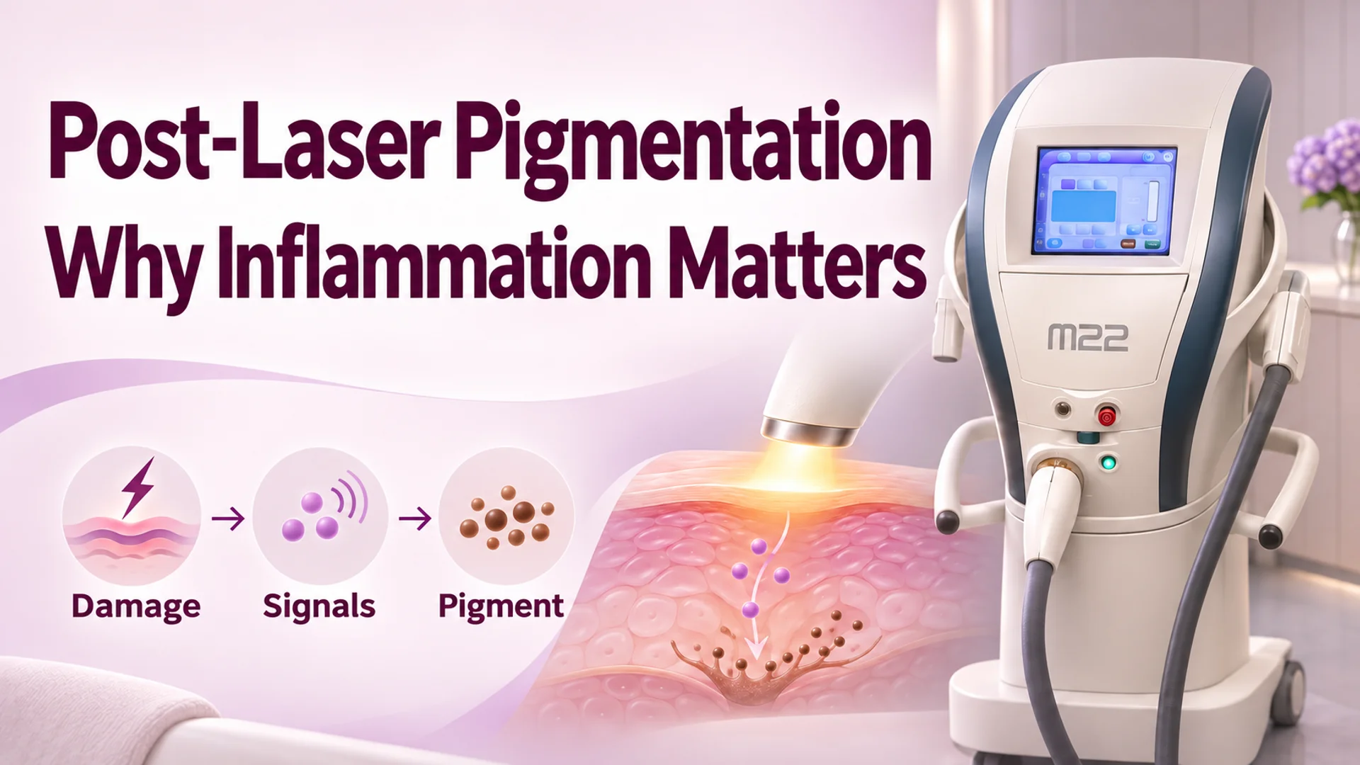
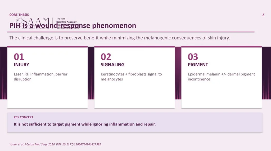
The issue is that whether it is a nanosecond laser or IPL, an unavoidable physiological response occurs during the process of applying heat or light energy to destroy melanin pigments in the skin. When the skin experiences heat or physical damage, skin cells (keratinocytes) activate a sort of defense mechanism. In 2026, at the SAAM conference, Dr. Iskandar Vaisov from Uzbekistan explained this process as a paracrine cascade (intercellular signaling pathway between adjacent cells) and pointed out the biological limits of the skin 3.
When keratinocytes sensing heat damage release inflammatory signaling molecules such as Interleukin-1 beta (IL-1β) or Endothelin-1 (ET-1), adjacent melanocytes (pigment-producing cells) perceive them as stimuli. Inside the melanocytes, Microphthalmia-associated Transcription Factor (MITF) is activated, promoting the production of the TYR (tyrosinase) enzyme, which actually produces melanin 3. In other words, the more energy is applied to erase melasma, the more the skin judges that "it has suffered damage and must mass-produce defense substances," and instead synthesizes new pigment.
This response appears more strongly particularly in Fitzpatrick skin types III–IV (intermediate to dark skin tones with abundant melanin and sensitive inflammatory reactions). Dr. Vaisov warned that on such skin, "targeting only pigment and ignoring inflammation and the recovery process is not sufficient," and that repeated overtreatment can worsen pigmentation 3.
How Combination Therapy Stabilizes 'Late-Stage Indicators'
The core reason combination therapy is gaining attention is not because it shoots energy more intensely to destroy more pigment. It is because the combined medication controls the secondary pathway of 'heat damage → inflammation → new pigment production' that unavoidably occurs during the procedure.
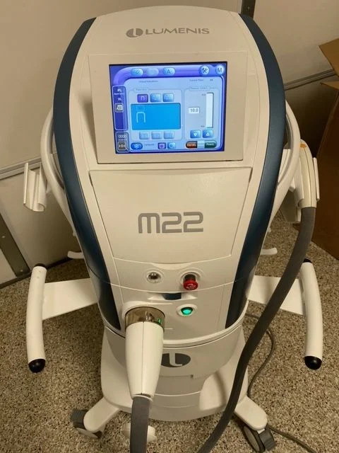
The results of this combination strategy were specifically demonstrated in the five-month follow-up RCT by the Shenyang China Medical University research team. Thirty-three adult patients with Fitzpatrick skin types III–IV received four sessions of IPL (Lumenis M22, 13–17 J/cm²) at four-week intervals. One group applied a 3% tranexamic acid hydrogel patch to the melasma areas for 20 minutes every evening, while the other group used a placebo patch without drugs 1.
Initially, the melasma severity (mMASI) scores improved in both groups, but a divergence appeared in the precise measurement indicators.
- Month 1 (Initial Inflammatory Stage): The IPL-only placebo group showed a +1.6% increase in melanin index. The authors interpreted this as a hidden Post-Inflammatory Hyperpigmentation (PIH) response due to the thermal stimulation of IPL. Conversely, the tranexamic acid combination group maintained a downward curve with a -7.8% decrease in melanin index 1.
- Month 5 (Stabilization Stage): After five months, melasma severity decreased by 50.5% in the combination group, while it decreased by only 38.5% in the single-treatment group. Particularly, the improvement in melanin index was overwhelmingly larger in the combination group (-36.3%) compared to the single-treatment group (-1.3%) 1.
The specific mechanism by which tranexamic acid directly 'physically blocks' the thermal signals of IPL is still suggested at the level of the authors' views. The authors explain that the anti-inflammatory effects of tranexamic likely suppressed the overall inflammatory response induced by IPL. However, related to the result that scores for UV spots and skin texture significantly improved only in the combination group, they suggested the possibility that a decrease in mast cell numbers and suppression of Vascular Endothelial Growth Factor (VEGF) expression played a role 1.
This 'time lag' is also observed in broader medical evidence. According to a meta-analysis in the journal Medicine (2026) compiling 11 RCTs (total 461 patients), there was no significant difference between the combination therapy group and the single-treatment group at week 4 of treatment. However, by weeks 8 to 16, the improvement in MASI for the combination therapy group was significantly greater than that of the single-treatment group by a Standardized Mean Difference (SMD) of −0.55 (95% CI −0.74 to −0.36) 4. The impact of initial stimulation appears to resolve over time, but the meta-analysis does not separately present the causal mechanism.
| Procedure Stage | Single Laser/Light-Based Group | Combination Therapy Group (Procedure + Topical Agent) |
|---|---|---|
| Week 4 (Initial | No significant difference in melasma severity (MASI) between groups | No significant difference in melasma severity (MASI) between groups |
| Weeks 8–16 (Late | Less improvement compared to the combination group | Additional improvement of SMD −0.55 compared to single group (statistically significant, 95% CI −0.74 to −0.36) |
The Medicine (2026) meta-analysis reported that while there was no advantage of combination over single therapy at week 4, the decrease in melasma severity in the combination group was significantly greater after week 8 4.
Ultimately, combination therapy is a strategy that stabilizes the downward curve of late-stage indicators by suppressing the pathway where the skin produces pigment again due to procedures intended to erase it, rather than a method that increases the immediate speed of pigment destruction right after treatment.
Limitations of Evidence and Points of Caution
While it is true that combination therapy is more favorable for long-term improvement than single therapy, it is not a universal solution applied equally to all patients and all devices. The most important thing is that medications combined with treatment can burden the skin. In the meta-analysis of 11 RCTs, the risk of side effects such as erythema (redness) or PIH (pigmentation) was statistically significantly higher in the combination therapy group than in the single-treatment group (Odds Ratio 8.96) 4.
However, the certainty of evidence for safety data in the meta-analysis was rated as 'very low,' so the reliability of the side effect frequency itself is limited 4. On the other hand, in the RCT combining IPL and a 3% tranexamic acid topical patch, which was covered as the core case in this article, there were no clinical adverse events in either group 1. While there may be specific combinations where you do not need to worry about side effects, caution is required as it is not unconditionally safe.
Additionally, currently verified strong clinical evidence is mainly concentrated on protocols combining IPL and tranexamic acid, and five-month follow-up studies targeting patients with Fitzpatrick skin types III–IV 4, 1. Large-scale RCTs directly comparing the exclusive use of the latest nanosecond/picosecond lasers with combination therapy are not included in the data verified this time. However, the conceptual framework presented in Dr. Vaisov’s lecture—'Energy damage → Keratinocyte signal → Melanocyte stimulation'—is a general principle applicable to energy-based procedures in general, so it is difficult to view the latest nanosecond lasers as completely an exception to the same inflammation and pigment recharge pathway.
Energy-based treatments are excellent at destroying melasma pigment, but they cannot avoid the skin's defense response of synthesizing new pigment due to the heat and inflammation generated in the process. Combination therapy is gaining attention not because the latest devices cannot erase pigment, but because the improvement in MASI in the combination therapy group was significantly greater by an average of 0.55 than in the single-treatment group at weeks 8–16 4. 1 The study authors interpret that the anti-inflammatory effects of the combined tranexamic acid likely suppressed the inflammation induced by IPL and the subsequent pigment recharge pathway, but the mechanism itself remains at the level of the authors' proposal 1.
References
- Эффективность и безопасность комбинированной терапии интенсивным…: рки | Атлас косметолога · xn--80ajoijsj.xn--p1ai↩
- Hyperpigmentation: Types, Causes and the Treatment Ladder · acibademinternational.com↩
- SAAM_2026_PIH_Deck - online presentation · en.ppt-online.org↩
- Пигментация и мелазма: исследования на русском | Атлас косметолога · atlas-kosmetologa.ru↩